How a Vet Diagnoses and Treats Cancer in His Own Dog
So, my dog Cardiff has cancer. My own pooch, who has overcome three bouts of Immune Mediated Hemolytic Anemia (IMHA) in his nearly nine years of life now has a fatal disease. If you’re first reading of this, I started the chronicle of Cardiff’s cancer journey in my last petMD Daily Vet article, Can a Veterinarian Treat His Own Pet?
Dr. Schochet of Southern California Veterinary Imaging (SCVI) discovered Cardiff's intestinal mass via ultrasound. Unfortunately, the ultrasound diagnosis doesn't determine the exact cellular nature of the mass. The suspicion was high for Cardiff’s mass to be cancer, but based on his lack of severe clinical signs and the appearance of the affected site on his abdominal ultrasound, the potential existed for Cardiff to not have cancer; granuloma was still a possibility. Granuloma is an area of inflammation typically caused by the body’s response to a piece of embedded foreign material or localized area of infection (bacteria, virus, parasite, etc.).
Biopsy would clarify this quandary. If Cardiff did have cancer, then the biopsy would also determine if the cells were benign (less concerning) or malignant (more concerning).
Attaining a fine needle aspirate for cytology (microscopic evaluation of cells) or biopsy via ultrasound was’t happening due to the challenging location of the mass deep within Cardiff’s abdomen. So, surgery was needed to remove the mass. The great news about surgery is that it also could potentially be curative. Additionally, the exact nature of his illness could be determined via biopsy so that the most-appropriate, post-surgical treatment could be started.
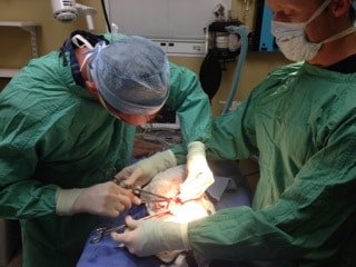
My veterinary associate, Dr. Mark Hiebert, performed the surgery with my assist. Having neutered Cardiff as a puppy, I feel comfortable performing surgery on him but I’m somewhat out of practice when it comes to major abdominal procedures.
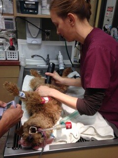
Cardiff’s vital organs were working perfectly, so he was an ideal anesthetic candidate. One of my most-trusted veterinary technicians, Dawn McCoy, was also on hand to oversee the anesthetic induction, maintenance, and recovery process. So, I had confidence that Cardiff would sail through his surgery with flying colors.
Upon opening Cardiff’s abdomen I was relieved to not see any obvious evidence of disease in his other abdominal organs but for the discrete mass on his jejunum (middle portion of his small intestine). Cardiff underwent intestinal resection and anastomosis, which means we removed an unhealthy section of his intestine (with wide margins) and then put back together the two healthy-appearing free ends.
The small intestine is held together by a fibrous netting of tissue called the mesentery, which contain lymph nodes that drain the intestines. As disease from one area of the intestines can spread to other parts of the body through the lymphatic system, it's vital to biopsy the mesenteric lymph node adjacent to the surgery site to determine if disease was already spreading. Fortunately, the lymph node that was biopsied visually appeared normal.
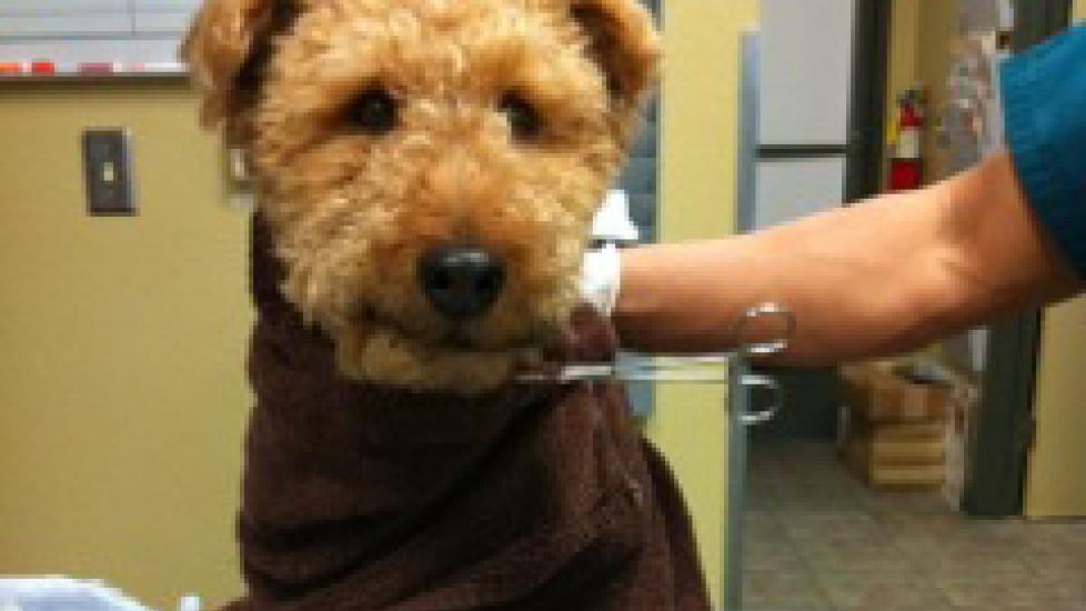
Cardiff had an uneventful anesthetic recovery, which I promptly "photo-bombed" for commemorative sake. Once his endotracheal tube was removed, he began to look like a much more normal, yet drugged, version of himself. To ensure his continued positive recovery, Cardiff spent the night in the hospital so that he could receive intravenous fluids, antibiotics, and pain medication.
While waiting with bated breath for the biopsy results, I was still hopeful that there could be a chance that Cardiff may not have cancer at all. If Cardiff had a granuloma instead of cancer, then surgical removal would be curative.
Unfortunately, Cardiff’s biopsy did not show a granuloma. Cardiff was instead diagnosed with a severe form of cancer that may shorten his lifespan, especially if it were to go untreated via surgery or chemotherapy.
Cardiff was diagnosed with “transmural malignant round cell sarcoma with mesenteric invasion, consistent with high-grade malignant lymphoma.” Lymphoma is white blood cell cancer. Either B or T cell lymphoma could be the cause of Cardiff’s mass, so immunophenotype staining of the tissue was required to differentiate between the two types of lymphoma. Just to keep the suspense building, the results of the test would take 10 to 14 days to process.
On a positive note, the mesenteric lymph node showed no evidence of cancer. There was evidence of inflammation associated with the tissue changes that were occurring at the site of the mass, but to my relief the cancer had not spread further.
Cardiff is healing well from his surgery and will start a course of chemotherapy in the coming weeks. Never a dull day for this veterinarian and his canine companion!

Dawn McCoy prepares Cardiff for surgery

Drs. Mark Hierbert (L) and Patrick Mahaney (R) perform Cardiff’s cancer surgery

Dr. Mahaney photo-bombs Cardiff post-surgery

Dr. Patrick Mahaney
