5 Cool Tech Advances in Surgery for Pets

By Paula Fitzsimmons
Your beloved pet needs surgery and you’re understandably concerned about putting her through a risky procedure and lengthy hospital stay. The good news is that technology is helping make the process safer and less stressful for animals.
In the last few years, new advances in veterinary technology have improved the way vets diagnose, treat, and manage diseases, says Dr. Cassie Lux, an assistant professor at the University of Tennessee, College of Veterinary Medicine in Knoxville.
Here is a look at some of the developments helping to improve the lives of our canine and feline companions.
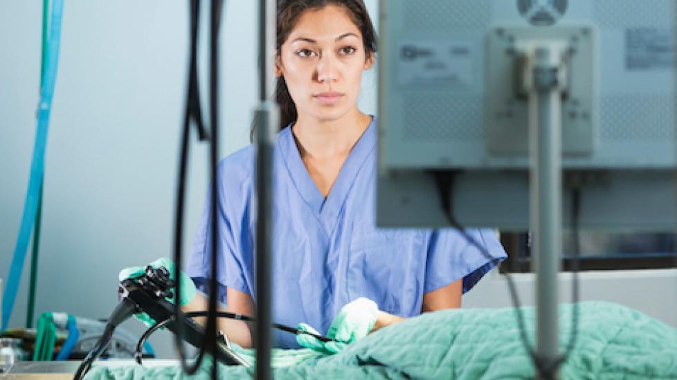
1. Flexible Endoscopy
The ability to perform minimally-invasive interventions of the gastrointestinal tract, urinary tract, and airways is one of the most widely available tech advances in veterinary care, says Lux, who is board certified in veterinary surgery.
Vets use an endoscope, a fiber-optic device that takes pictures of internal organs, magnifies the images, and displays them on high-definition medical monitors. “This technology provides an excellent viewing field in areas that can be traditionally very difficult to visualize,” Lux says.
Depending on the type of procedure being performed, endoscopes can be rigid or flexible. “With flexible endoscopy of the airways, gastrointestinal tract, and urinary tract, diagnoses for conditions and treatments can be performed without any need for incisions,” she says. The benefits are reduced pain and a speedier recovery time for the animal.
Examples of procedures using flexible endoscopes include removal of ingested or inhaled foreign objects, treatment of urinary stone disease, and biopsy procurement for GI and urinary diseases, she says.
2. Rigid Endoscopy
Rigid endoscopes allow vets to perform minimally-invasive procedures of non-tubular regions like the abdominal cavity (laparoscopy) and thoracic cavity (thoracoscopy), Lux says.
A major advantage of rigid endoscopy is that unlike traditional surgery, vets only need to make small incisions to get the same results. For example, in a procedure to remove ovaries, the surgeon makes two 5 millimeter incisions, says Dr. Kathleen Ham, an assistant professor at the Ohio State University, College of Veterinary Medicine in Columbus, versus a big abdominal incision that is required for traditional surgery.
“This can be beneficial in any patient, but imagine the benefit in older dogs, obese dogs, and dogs in heat that require bigger incisions and may be prone to more complications,” Ham says. “Patients are up and moving pretty quickly after surgery and owners are happy to be able to provide a surgical option similar to what they would receive.”
Minimally-invasive surgery reduces the amount of tissue trauma and pain associated with surgery, says Ham, who is board certified in veterinary surgery. “You also get many other benefits, such as reduced bleeding and enhanced visualization with magnification and illumination, and you can easily record and take pictures for documentation.”
Most traditional (open surgery) procedures now offer minimally-invasive options. Some of these include multiple abdominal organ biopsies, gallbladder removal, abdominal testicle removal, spay procedures, and lung biopsy, Lux says.
3. Interventional Radiology
Interventional radiology is a relatively new specialty that has gained widespread interest in recent years, Lux says. The equipment is similar to what human doctors use, “including long diagnostic catheters, guidewires to access vascular paths or openings, devices used to form blood clots, balloon catheters to open narrowed or stenotic regions, and stents of various composition to maintain the shape of, widen, or hold open a vessel or part of an organ.” (Vets use tubular devices called stents to keep blocked passageways open.)
The technique is either performed via passages like the mouth, airway, or urethra, or through blood vessels (either through the groin or neck), she says.
According to Dr. Lynetta Freeman, an associate professor at Purdue University College of Veterinary Medicine in Lafayette, Indiana, a number of conditions are now treated with interventional radiology. These include “delivery of tracheal stents to hold open the airway for dogs with tracheal collapse, or animals with a tracheal stricture; delivery of an occlusion device to block a PDA (patent ductus arteriosus), a vessel that fails to close after birth and results in abnormal blood flow; delivery of stents that alleviate obstructions in flow of urine (kidney to bladder, bladder to urethra); delivery of coils and/or embolic agents that block blood flow to a tumor to reduce its growth; and targeted delivery of chemotherapy agents directly into the blood vessel supplying a tumor.”
The primary benefit of interventional radiology is that it reduces the level of invasiveness, as compared to traditional surgical procedures, says Freeman, who is board certified in veterinary surgery. “The technique can also address conditions that we previously thought were hopeless, offering owners an opportunity for palliative care for their pets.”
Another pro is that it reduces down time, Freeman adds. “Although these procedures are performed under general anesthesia, many times animals receiving this approach are able to go home the same day, as compared to a lengthy hospitalization.”
4. Surgical 3D Printing
Unlike X-rays, which provide two-dimensional views, 3D printing creates a realistic, tangible model. “The utility of 3D printing for visualization of disease conditions prior to surgical procedures greatly improves the surgeon’s ability to understand all aspects of treatment, and formulate a complete plan in a lower stress environment than an operating room,” Lux says.
The process begins with computerized axial tomography (also known as a CAT scan or CT scan), which takes cross-sectional images of the patient, then transmits them to a monitor. Information from the scan is used to make or draw a bone, says Dr. Robert Hart, director of orthopedic and joint replacement surgery at Animal Medical Center in New York City. Veterinarians can then use a computer mouse to spin or twist the bone on their screen to get a better idea of what’s happening to the bone and what level of deformity it has, he says.
Hart recently treated a 7-month-old Irish Setter whose limb was deformed at different angles. The dog wasn’t in any pain, but because the leg was so deformed, he walked with an awkward gait and was at risk for early arthritis. After taking an X-ray, which provided minimal information, he ordered a CT scan and sent it to an outside company, which put it through a 3D printer and created a model of the dog’s leg. “It’s fit-to-scale, made of a resin-like plastic that simulates the hardness and the texture, the way a bone feels…so we can hold it in our hands and study it even further,” he describes.
This technology allowed Hart to practice his technique prior to the surgery. “We were able to make the cuts and study whether we were making them in the right places, and determine what the effects of the cut in the bone were,” says Hart, a board-certified veterinary surgeon who specializes in orthopedic surgery and joint replacement. “We could actually test the hardware we were going to use in surgery to hold the bone in the new or normal position.”
Rather than go into surgery blind and try to figure out how to straighten the bone, Hart had solved the problem in advance. This made the surgery faster and more efficient, he says. “And faster surgery is safer for anesthesia because the dog is under for a shorter period of time. It’s shorter for infection rates because the longer the dog is under anesthesia, the higher the potential there is for infection.”
5. Laser Therapy
Laser therapy is one of the most versatile tools in a veterinarian’s toolbox, says Maria C. Caiozzo, a certified canine rehabilitation technician. Low-level lasers (also known as cold lasers) transmit wavelengths of 800 to 900 nanometers, which she says impart a myriad of benefits for animals.
These include “a reduction in pain and inflammation, increased circulation to promote the healing process after injuries or surgery, and improved mobility for more functional strengthening to get animals back on their feet faster post-surgery, leading to fewer long-term complications,” says Caiozzo, a client growth consultant at Respond Systems and RSI Equine.
Laser therapy is used in a variety of procedures, including dental extractions, spays and neuters, soft tissue surgeries, wound healing, and the management of chronic pain and inflammatory conditions, she says.
“With animals living longer, just like their humans, the veterinary rehabilitation market is booming and practitioners are looking for new technologies to manage and treat conditions that affect animals in their older age,” Caiozzo says. “Technologies to not only treat chronic inflammatory conditions in a pet’s senior years, but also help prevent them through increasing the effectiveness of PT and rehab in an animal’s younger years.
“Laser therapy is the most widely used modality in rehabilitation and sports medicine practices across the country and internationally,” she adds, “and will continue to be an integral part of healing pets as this industry continues to grow.”